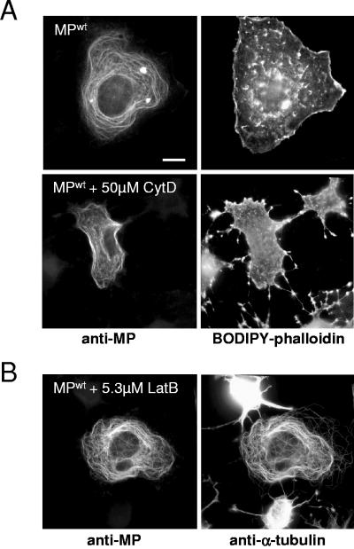
FIG. 5.
MP does not colocalize with actin. (A) MPwt-transfected cells were stained with antibodies specific for MP and with BODIPY-558/568-conjugated phalloidin for actin. MP shows microtubule-specific distribution (top left) and no spatial overlap with actin (top right). Treatment of cells with cytochalasin D affects the pattern and function of actin (bottom right) without changing the circular-filamentous pattern of MP distribution (bottom left), indicating that this distribution is actin independent. (B) MPwt-transfected cells treated with latrunculin B and stained with antibodies for MP (left) and α-tubulin (right). Microfilament disruption is evident in untransfected cells, which show clear effects on cell shape (right, top and bottom cells). In MPwt-transfected cells, both cell shape and the association of MPwt with microtubules are not affected. Bar = 10 μm.

