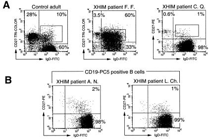
Figure 2.
Fluorescence analysis and sorting of IgD+CD27+ peripheral blood B cells of XHIM patients and a control adult donor. (A) Anti-IgD/anti-CD27 two-color staining of CD19+ B cells enriched by magnetic cell sorting (MACS) was performed as described in Materials and Methods. The gates selected for sorting of the IgD+CD27+ B cell population are indicated. The IgD−CD27+ population present in XHIM patients corresponds to a residual T cell contamination (see B and Materials and Methods). The sorted fractions were used for sequence analysis of rearranged VH3–23 gene segments. (B) Anti-IgD/anti-CD27/anti-CD19 three-color staining performed on peripheral blood mononuclear cells confirmed the absence of IgD−CD27+ memory B cells in XHIM patients. Three-color analysis shows CD27 and IgD expression on CD19+-gated cells. The data are representative of all XHIM patients studied.

