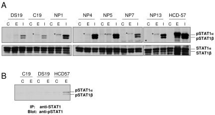
FIG. 4.
Analysis of tyrosine phosphorylation of STAT1 in SFFV-transformed erythroleukemia cell lines. (A) SFFV-transformed erythroleukemia cells were left unstimulated (C) or stimulated with Epo (100 U/ml) (E) or IFN-α (500 U/ml) (I) for 15 min. Total cell lysates were then immunoblotted with either anti-phospho-STAT1 (tyrosine 701) (upper panel) or anti-STAT1 antibody (lower panel). Asterisks indicate nonspecific bands. HCD-57 cells (far right), derived from an F-MuLV-infected mouse, were used as a positive control. (B) To demonstrate that the nonspecific band marked by an asterisk in panel A was not STAT1, SFFV-transformed C19 and DS19 cells were left unstimulated (C) or stimulated with Epo (100 U/ml) (E) for 15 min, and then total cell lysates were immunoprecipitated with anti-STAT1 antibody. The immunoprecipitated proteins were then separated by electrophoresis, transferred to nitrocellulose, and immunoblotted with anti-phospho-STAT1 antibody. HCD-57 cells were used as a positive control.

