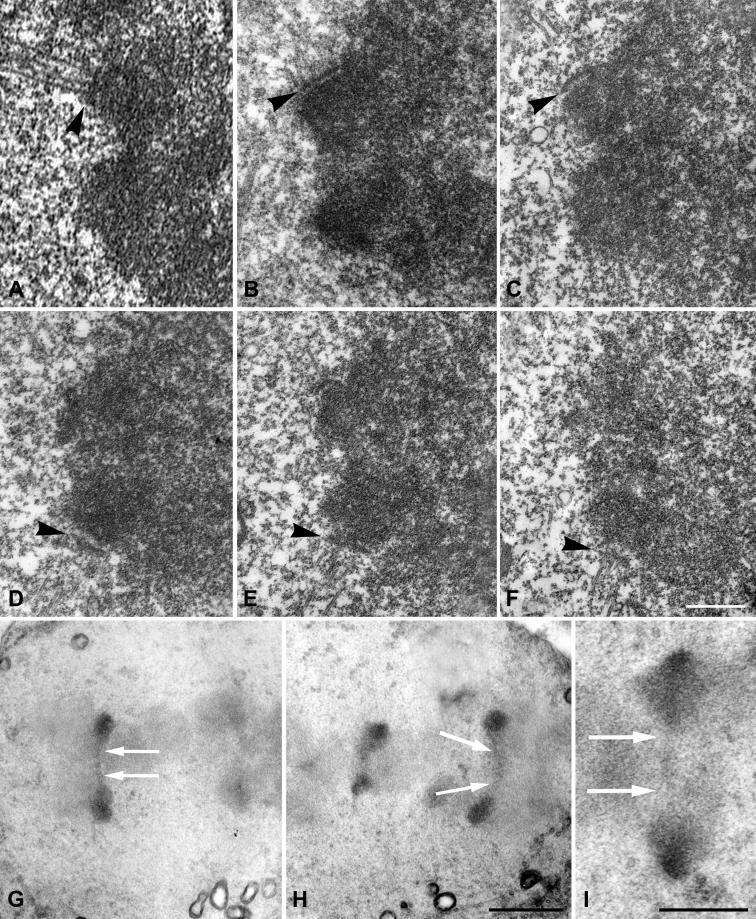
Figure 6. Ultrastructure of Metaphase II Centromeres.
(A–F) Six serial sections of a metaphase II centromere. Conventional technique. The outer plates of both sister kinetochores (arrowheads) are clearly differentiated.
(G and H) Two sections of metaphase II centromeres after the Os-PPD technique. The condensed chromosomes show a very low contrast, while sister kinetochores show a high contrast. The kinetochores appear joined by a continuous band of medium contrast material (arrows).
(I) Centromeric region of a chromosome in late metaphase II. A low contrast material extends as a thin and discontinuous band (arrows) joining the highly contrasted sister kinetochores.
Bars, 0.5 μm (A–F), 2.5 μm (G,H), and 2.5 μm (I).

