Figure 4.
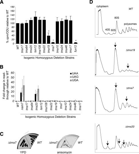
Deletions of TMA proteins result in translation defects. (A) In vivo translation assay. Translation was measured as the amount of 35S-methionine incorporation in yeast strains deleted for the indicated TMA protein relative to the isogenic wild-type (WT) strain. The standard deviations between three independent samples are shown. (*) A p-value of <0.05. (B) Nonsense suppression assay. Translation of a β-gal reporter gene containing either UAA, UAG, or UGA mutations, in deletion strains relative to the wild-type strain (set as 1). The standard deviations from at least three independent experiments with three distinct colonies for each strain are shown. (C) Resistance to translation inhibitors. YPD or YPD + 50 μg/mL anisomycin plates streaked with wild-type or Δtma7 after incubation for 2 d at 30°C are shown. (D) Polysome profile analysis. Absorbance at 254 nm of sucrose gradient fractions from wild type, Δtma19, Δtma7, and Δtma20. Arrows indicate differences in the mutant profiles compared with wild type.

