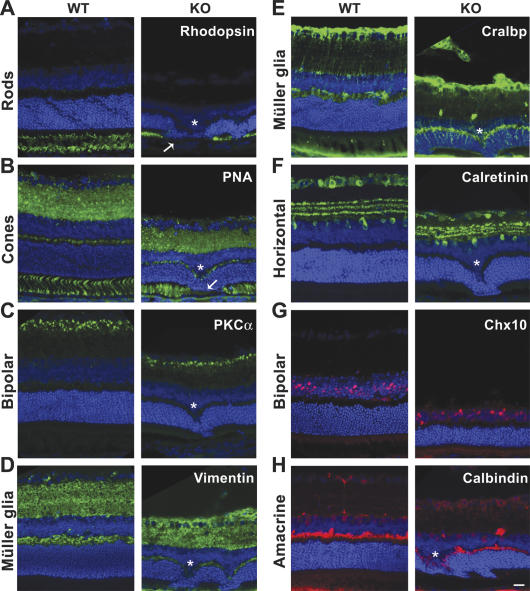
Figure 3.
Normal cell-type specification in Tlx−/− retina. Cryosections of neural retinas from E16.5 to P21 mice were stained for cell-type-specific markers (red or green) by immunohistochemistry. The sections were then counterstained for nuclei to reveal the nuclear layers (blue). Typical images from P21 sections are shown. (A,B) Immunohistochemistry for rods (rhodopsin) and cones (PNA). Asterisks indicate the protrusions from inner nuclear layer. Please note the discontinuous staining for rods and cones (arrows) around the protrusions. (C,G) PKCα- and Chx10-positive bipolar cells. (D,E) Vimentin- and Cralbp-positive Müller glia. (F,H) Calretinin- and calbindin-positive horizontal cells. (H) Calbindin-positive amacrine cells.

