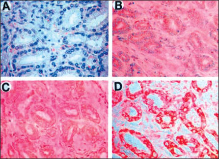
Figure 10.
Four different prostate cancers stained with a monoclonal antibody for Type 1 5AR (red color), showing that in some cancers (ie, section D above), the enzyme is highly expressed. Marginal expression of the enzyme is seen in cancer A, moderate expression in cancers B & C. 5AR, 5α-reductase. Reprinted with permission from Thomas LN et al.32

