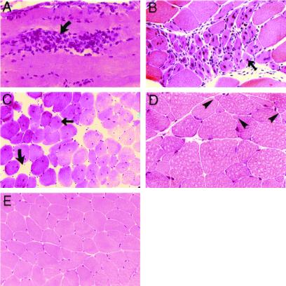
Figure 2.
Muscle histology: focal areas of degeneration, regeneration, and variation in fiber size. Hematoxylin and eosin-stained sections of the gastrocnemius (A), triceps brachii (B), paraspinal (C), and quadriceps (D) muscles of 6-month-old null mice showing cell degeneration (curved arrows, A and C) with macrophage infiltration (A), regenerative fibers (thin arrow, B), central nuclei (thin arrow, C), and increased variation in fiber size with atrophic muscle fibers (D, arrowheads) compared with age-matched wild-type mice (E). Original magnifications: A and D, ×300; B, ×200; C and E, ×80.

