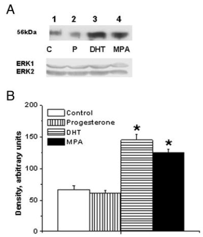
Fig. 5.

Effects of MPA and DHT on TP receptor expression in male rhesus coronary VMC. A, TP receptors in primary coronary VMC from gonadally intact, male RM treated for 72 h as follows: C, untreated control (lane 1); P, P treatment (1 nm; lane 2); DHT, DHT treatment (10 nm; lane 3); and MPA, MPA treatment (10 nm; lane 4). ERK1/2 detection was used as an internal loading control. B, Quantitation of TP receptor expression in RM coronary VMC was performed by densitometric analysis using NIH-Scion Image analysis software. MPA and DHT treatments increased the expression of TP receptor, whereas P did not significantly change the expression of TP receptor. Data are expressed as the mean ± sem and represent results from two independent experiments performed in duplicate. *, Significant increase compared with respective untreated control coronary VMC (P < 0.05).
