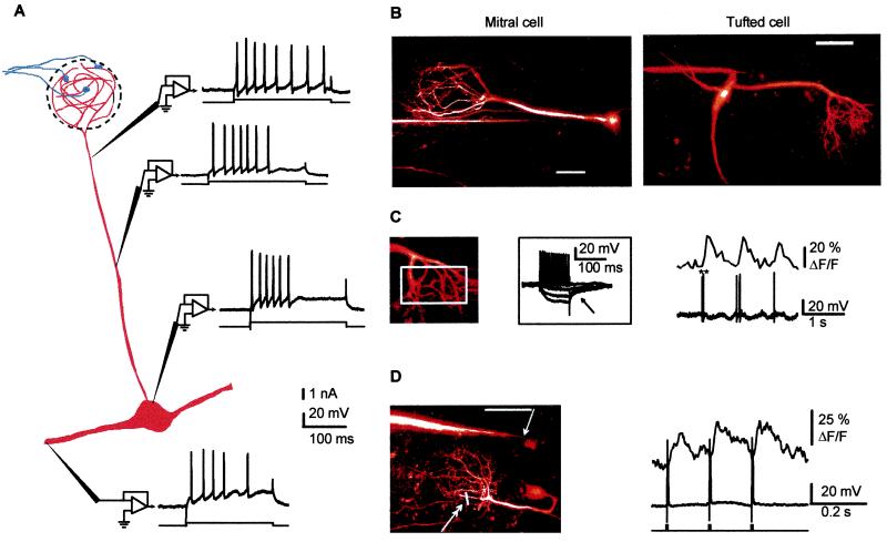
Figure 1.
Sodium action potentials synchronize [Ca2+] transients in all dendritic compartments of mitral cells in the olfactory bulb of anesthetized rats. (A) Fast sodium action potentials were observed with sharp electrode recordings from apical secondary dendrites and from the soma. Shown is a schematic diagram of mitral cell synaptic connections and responses from four different mitral cells to depolarizing current pulses. Sodium action potentials were seen at all sites. Note that the resting membrane potential and the injected currents varied slightly at each site. (B) Examples of mitral and tufted cells that were filled with Ca2+-Green-1 and imaged using two-photon microscopy (see Methods for recording and imaging techniques). Both pictures are two-dimensional projections of image stacks (up to 200 frames each separated by a 2-μm step in depth) obtained at the end of the recording session (scale bars = 50 μm). Electrophysiological recordings were obtained from the soma (Left, mitral cell, length of the apical dendrite, 210 μm) and at the origin of a secondary dendrite (Right, tufted cell, length of the apical dendrite, 130 μm). (C) Spontaneous action potentials induce a [Ca2+] rise in the tuft. (Left) Fluorescence signals indicative of [Ca2+] changes were integrated in the region indicated by a white rectangle (same tufted cell as in B; the ** indicates a spike doublet). The Inset Illustrates the firing of the cell for which hyperpolarizing pulses activated an IA-type current (arrow). (D) Action potentials propagate backward in vivo. (Left) A recording micropipette was placed in the secondary dendrite (arrow) of a mitral cell with a long apical dendrite (230 μm, scale bar = 50 μm), and fluorescence signals were measured in two tuft branchlets (double arrow) by using a line scan. (Right) Three single spikes, evoked with depolarizing current pulses (1.5 nA, 5 ms) injected in the secondary dendrite, induce fast [Ca2+] transients in the tuft. Because of the location of the current injection site distal to the soma, it is most likely that sodium action potentials were initiated in the secondary dendrite or possibly in the soma and propagated backward to the tuft (i.e., they did not initiate in the tuft dendrite). Note the rapid decay of the transient elicited by a single action potential.

