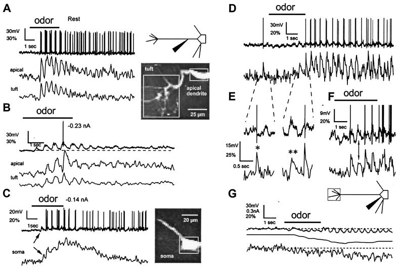
Figure 2.
Odor stimulation evokes two types of [Ca2+] changes in mitral cell dendrites. (A–C) Fluorescence measurements from the distal tuft, the apical dendrite at the edge of the glomerulus (200 μm from the soma), and the soma of a cell. The microelectrode was located in the apical dendrite 75 μm distal to the soma. (A Left) During odor stimulation (3 s isoamyl acetate), transient [Ca2+] increases were observed, phase-locked with bursts of action potentials that occurred during each respiratory cycle. [Ca2+] increases recovered substantially between breaths. (B) Subthreshold depolarizations evoke [Ca2+] changes. Hyperpolarization of membrane potential below the threshold for spike generation revealed underlying cyclical depolarizations during odor presentation. In both the apical tuft and dendrite, subthreshold slow depolarizations were co-incident with increases in [Ca2+]. Dashed line below voltage trace highlights the sustained depolarization after odor offset. (C) Increased [Ca2+] during odor-evoked subthreshold depolarization was also seen in the soma (same cell, apical dendrite projecting toward left and top). Vertical arrow indicates an increase in [Ca2+], which begins with the first subthreshold depolarization (fluorescence changes expressed as ΔF/F%). (D–G) Voltage dependency of depolarization- and action potential-evoked [Ca2+] increases in fine tuft dendrites. The microelectrode was located in the soma of another cell. (D) The mixed response to application of the odor was initially inhibitory overall but became excitatory toward the end of application with one subthreshold depolarization inducing a slow [Ca2+] increase (see below). (E) Expanded portion of records in D as indicated shows the rapid recovery kinetics of Δ[Ca2+] provoked by action potentials (∗) compared with subthreshold voltage depolarizations (∗∗). (F) Another odor application in the same cell. (G) A large hyperpolarization blocks all [Ca2+] changes. Hyperpolarizing current (middle trace) was progressively injected to oppose the increasing odor-induced excitation. [Ca2+] increases in phase with respiration-linked slow depolarizations were no longer observed. Instead, a reduction in the steady-state [Ca2+] level occurred.

