
FIG. 2.
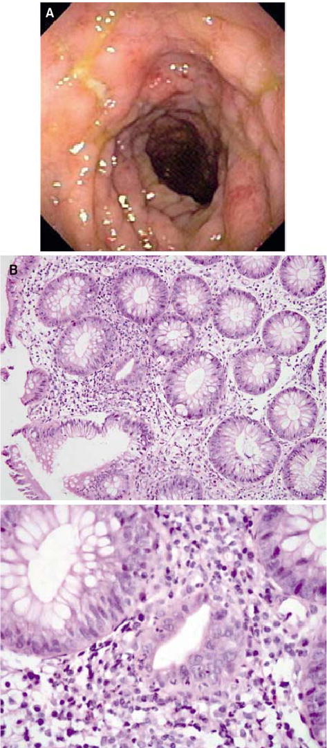
(A) Colonoscopic view of bowel edema and ulceration in the descending colon of patient 29, who experienced autoimmune colitis. (B) Histopathologic analyses revealed focal active colitis (upper panel) with crypt destruction, loss of goblet cells, and neutrophilic infiltrates in the crypt epithelium (lower panel) (original magnification: upper panel, ×20; lower panel, ×60).