
FIGURE 2.
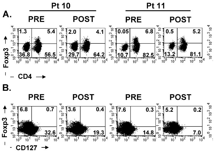
Peripheral blood mononuclear cells from 3 patients (60 λμg/kg dose) (A) were stained with fluorescein isothiocynate-conjugated CD4, PerCP-conjugated CD3, and APC-conjugated CD25 antibodies followed by intracellular staining with biotinylated Foxp3 antibody. The dot plots were gated on CD3+ T lymphocytes. B, Peripheral blood mononuclear cells from these patients were also stained with fluorescein isothiocynate-conjugated CD4, PE-conjugated CD25, and antigen presenting cells-conjugated CD127 (IL-7Rαchain) followed by intracellular staining for Foxp3 protein. The dot plots were gated on CD3+CD4+ T cells. The quadrants were set based on isotype control antibodies as well as negative control, and the numbers represent the percentage of cells in each quadrant.