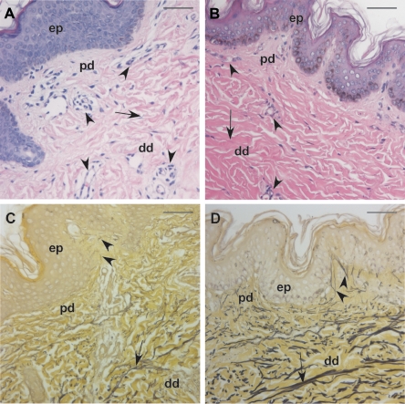
Figure 2.
Skin pathology in the proband. A and B, Hematoxylin-eosin staining of samples from the patient (A) and an age- and sex-matched control (B) revealed increased vascularization (arrowheads) and reduced collagen bundle size (arrows) in the dermis of the patient. C and D, Hart’s elastin staining showed severely underdeveloped elastic fibers in both the papillary dermis (pd) (arrowheads) and the deep dermis (dd) (arrows) of the patient (C) compared with a matched control (D). There is no apparent pathology in the epidermis (ep). Magnification bars = 50 μm.

