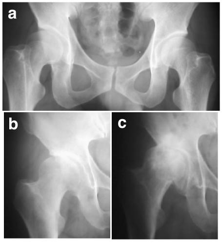
Figure 2. .
Hip changes in the study family. a, Radiograph of subject IV-5, at age 38 years, pre-OA. Radiological changes other than acetabular dysplasia are minimal; neither joint-space narrowing nor osteophyte is observed, and the shape of the femoral head is normal. b, Radiograph of subject III-8, at age 62 years, showing early-stage OA and acetabular dysplasia. c, Radiograph of subject III-8 at age 68 years, with advanced-stage OA.

