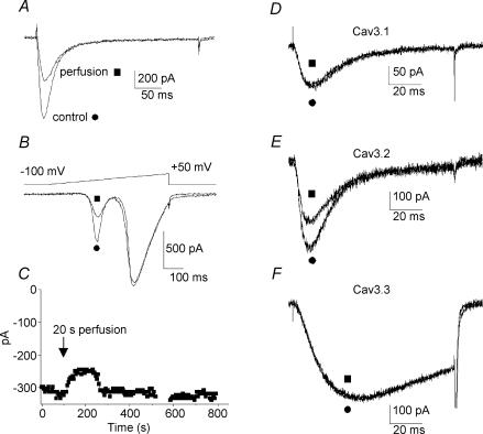
Figure 4. Mechanical responses of T-type current to the extracellular recording medium perfusion.
A, original trace of T-type current recorded in E17 vestibular neurone, before (•) and after (▪) application of extracellular recording medium at a velocity of 1 μl s−1. B, corresponding voltage ramps showing the absence of shift in I–V relationship. Note that HVA calcium currents were not reduced. C, long duration recordings showing the reversibility of the extracellular medium perfusion effect on vestibular neurones. Application of recording medium (20 μl, 20 s) induced a current decrease, which reached a minimum in about 60 s and was followed by an amplitude increase to return to the initial amplitude after about 220 s. D–F, T-currents recorded in HEK-293 cells transfected with the different Cav3/α1 T-channel subunits. Trace currents were illustrated before (•) and after (▪) application of extracellular recording medium at a velocity of 1 μl s−1. While Cav3.1/α1G (D) and Cav3.3/α1I (F) were insensitive to the perfusion, Cav3.2/α1H (E) decreased by about 30%.

