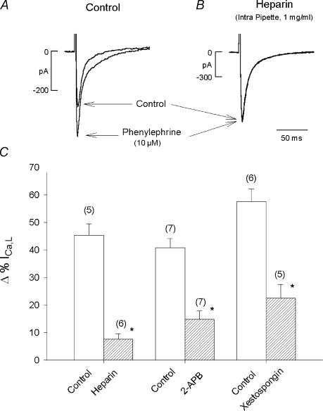
Figure 3. Inhibition of IP3 signalling inhibits PE-induced stimulation of ICa,L.
A, original records showing the effects of PE to stimulate ICa,L recorded with a ruptured patch method. PE elicited a typical increase in ICa,L amplitude. B, another atrial myocyte in which ICa,L was recorded during intracellular dialysis of heparin (1 mg ml−1) contained within the pipette solution. PE failed to increase ICa,L amplitude. In two additional series of experiments, PE was tested in the absence and presence of 2 μm 2-APB or 10 μm xestospongin C (incubation 2 h) using a perforated patch recording method. C, summary of the inhibitory effects of heparin, 2 μm 2-APB or 10 μm xestospongin C. Each IP3 receptor blocking agent significantly inhibited PE-induced stimulation of ICa,L. Numbers in parentheses indicate the number of cells tested in each experiment. *P < 0.05.

