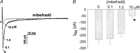
Figure 4. Inhibition of INa by mibefradil.
A, every 5 s, DVR pericytes were depolarized from a holding potential of −90 mV to −10 mV for 50 ms. During the pulses, increasing concentrations of mibefradil were introduced into the extracellular buffer. Superimposed traces show an example of the inhibition of INa as mibefradil was increased from 0 μm to 0.1, 1 and 10 μm. B, summary of peak inward INa versus mibefradil concentration. *P < 0.05 versus 0 μm mibefradil, n = 4, 6 and 10 cells at 0.1, 1.0 and 10 μm, respectively.

