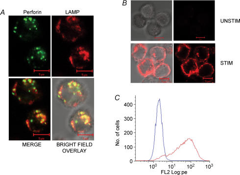
Figure 2. LAMP colocalizes with perforin in unstained cells and is exposed to the extracellular solution during stimulation.
A, colocalization between perforin and LAMP-1 in unstimulated cells. TALL-104 cells were fixed, permeabilized and stained with FITC-labelled antiperforin antibodies (shown in green) and PE-labelled anti-LAMP-1 antibodies (shown in red). Also shown are merge and bright-field overlay images in the bottom row. Yellow spots represent colocalized pixels. Scale bar is 5 μm. B, stimulation-dependent LAMP-1 staining in intact cells. In the presence of LAMP-1-PE antibody, TALL-104 cells were either incubated for 50 min in Normal Ringer solution (Unstim) or stimulated with TG + PMA (Stim). Shown are bright field and fluorescence images of both conditions. Scale bar is 5 μm. C, histograms of flow cytometry data showing an increase in LAMP-1 staining due to stimulation. TALL-104 cells were left untreated (blue trace) or stimulated with TG + PMA (red trace). Both were incubated in the presence of the LAMP-1 antibody.

