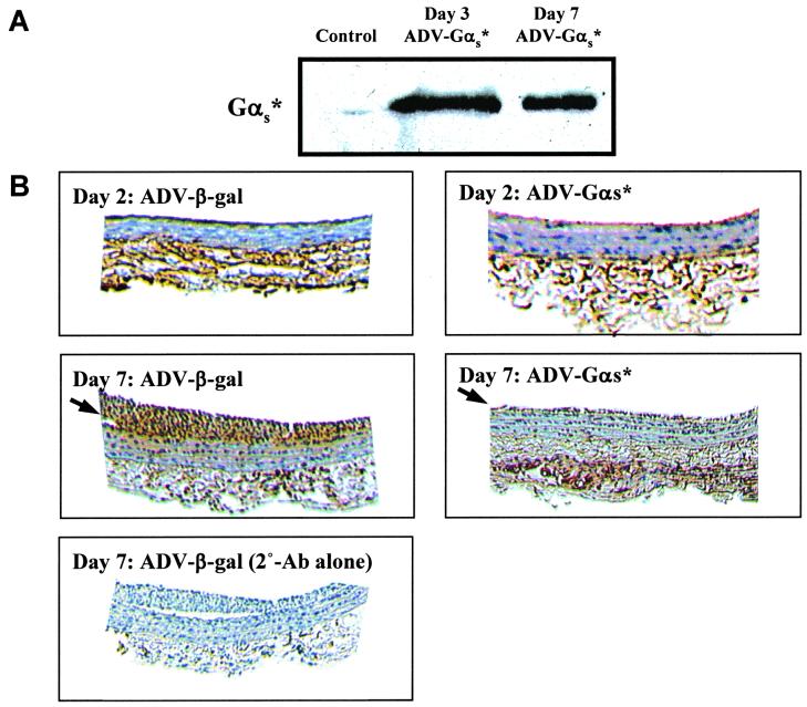
Figure 3.
Effect of activated Gαs expression on the MAPK activity in the carotid artery segments. (A) Immunoblot of injured carotid artery segments from control (uninfected) and αs*-infected animals. Arterial segments were homogenized in sample buffer and an equivalent amount of proteins from each sample was resolved by SDS-gel electrophoresis and blotted with the anti-FLAG m2 antibody. The 42 KDa region is shown. (B) Visualization of phospho-MAPK in the neointima of carotid arteries. Animals were subjected to balloon angioplasty and then infused with either ADV-β-gal or ADV-αs*. After 2 or 7 days, animals were killed, the carotid arteries excised, sectioned, and stained with phospho-MAPK antibody by using DAB as the chromogen (Top and Middle). As control, sections from ADV-β-gal-treated animals were stained without primary antibody (Bottom). Arrows indicate neointimal immunoreactivity.

