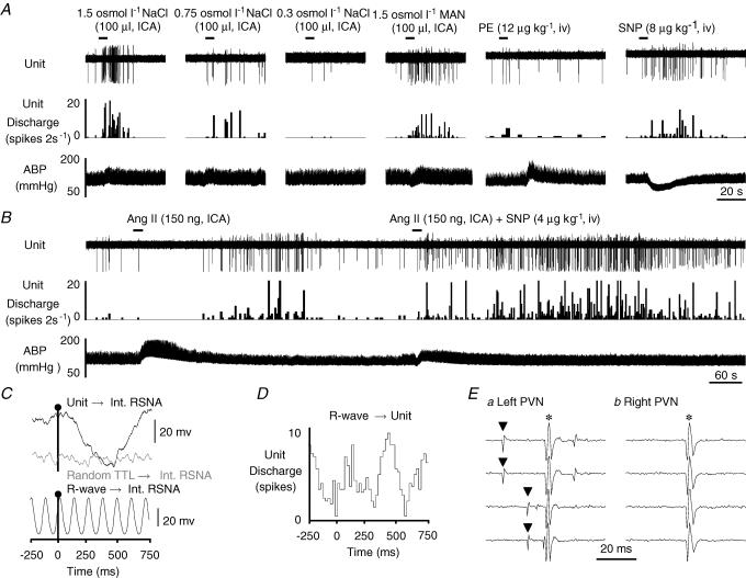
Figure 4. Example of a barosensitive type III MnPO-PVN neurone.
A, ICA injection of hypertonic NaCl dose-dependently increased MnPO-PVN cell discharge, whereas isotonic saline had no effect. Hypertonic mannitol (Man) also significantly increased the firing rate of this MnPO-PVN neurone. Moreover, this neurone was barosensitive as SNP decreased ABP and significantly increased cell discharge. B, ICA injection of Ang II produced a biphasic response with an initial and significant decrease in discharge rate when ABP was elevated followed by a significant increase in activity once ABP returned to baseline values. When the associated Ang II-evoked increase in ABP was attenuated with SNP, the neurone no longer showed a biphasic response. Instead, Ang II injection evoked an immediate and sustained increased in cell discharge thereby suggesting that the Ang II-evoked increase in ABP was masking the Ang II-excitatory effect on unit activity. C, baseline unit discharge significantly correlated with renal SNA (top trace, peak latency: 460 ms) whereas no obvious correlation was observed with randomly occurring square-wave pulses (random TTL→Int. RSNA, middle trace). The bottom trace shows that renal SNA is strongly correlated to the cardiac cycle during the same sampling time as revealed by R-wave-triggered averages of renal SNA. D, R-wave-triggered time histogram average of unit discharge showed a significant cardiac-cycle-related rhythm. The spike-triggered averages and histograms presented in C and D were constructed from 1366 spontaneous unit discharges. E, the neurone was antidromically activated from the left PVN with a constant onset latency of 14 ms and threshold of 130 μA (a, top two traces), followed high frequency stimulation (traces not shown), and the antidromic spike was collided with a spontaneous action potential (a, bottom two traces). In contrast, the same neurone could not be antidromically activated with a high stimulus intensity (1 mA, 1 ms) from the contralateral PVN (b). ▴, spontaneous action potential; *, stimulus artifact.

