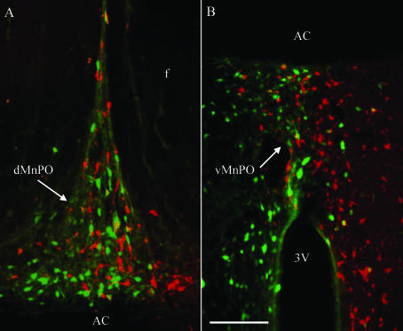
Figure 8. Example of retrogradely labelled neurones in the MnPO after microinjection of fluorogold and rhodamine-labelled microspheres into separate sides of the PVN.
Fluorogold-positive neurones are green, neurones with rhodamine-labelled microspheres are red, and neurones that co-localize fluorogold and rhodamine microspheres are yellow. Retrogradely labelled neurones from the PVN were present in both the dorsal (A) and ventral (B) MnPO as well as the precommissural MnPO. The number of retrogradely labelled neurones was not significantly different between retrograde tracers. Moreover, only 3.3 ± 1.0% and 3.8 ± 1.0% of fluorogold- and rhodamine-positive neurones, respectively, co-localized with the second retrograde tracer, thereby indicating that the majority of MnPO neurones have a unilateral projection to the PVN. Scal bar, 100 μm. dMnPO, dorsal median preoptic nucleus; vMnPO, ventral median preoptic nucleus; AC, anterior commissure; f, fornix; 3V, 3rd ventricle.

