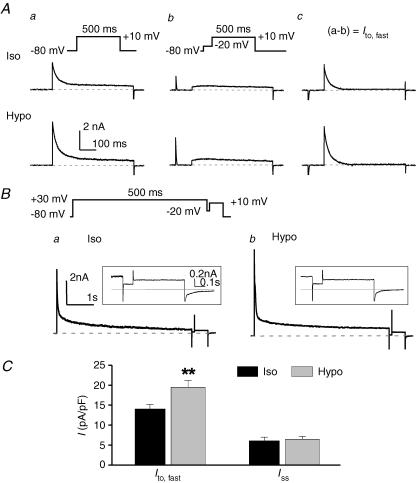
Figure 3. Hyposmotic cell swelling increases Ito,fast but not Iss in mouse left ventricular apex myocytes.
A, effects of osmotic stress on Ito,fast. Whole-cell currents were recorded in the presence of 50 μm 4-AP from the same cell with voltage protocols shown on the top under isosmotic (Iso, upper traces) and hyposmotic (Hypo, lower traces) conditions. Cells were held at −80 mV, and currents were elicited by a 500-ms depolarizing voltage pulse to +10 mV (a) or by a 500-ms depolarizing step preceded by a 100-ms ‘inactivating’ prepulse to −20 mV to inactivate Ito,fast.(b). Ito,fast was measured from the prepulse-sensitive difference currents obtained by subtracting the currents in panel b from the currents in panel a (a−b) under corresponding osmotic conditions (c). B, effects of hyposmotic cell swelling on Iss. The voltage protocol (shown on the top) consists of a 5-s, +30-mV step and a 0.75-s, +10-mV step, which is interposed by a 100-ms ‘inactivating’ prepulse at −20 mV. The membrane currents elicited by the depolarization step from −20 mV to +10 mV are Iss. Representative Iss recorded under isosmotic (Iso) and hyposmotic (Hypo) conditions are shown on expanded time and current scales in panels a and b. The dashed lines indicate zero current. Representative whole-cell current traces are shown in the inset. C, mean current densities of Ito,fast (Ac, n = 7) and Iss (B, n = 6) in mouse left ventricular apex myocytes under isosmotic (black bars) and hyposmotic (grey bars) conditions. **P < 0.01 versus isosmotic condition.

