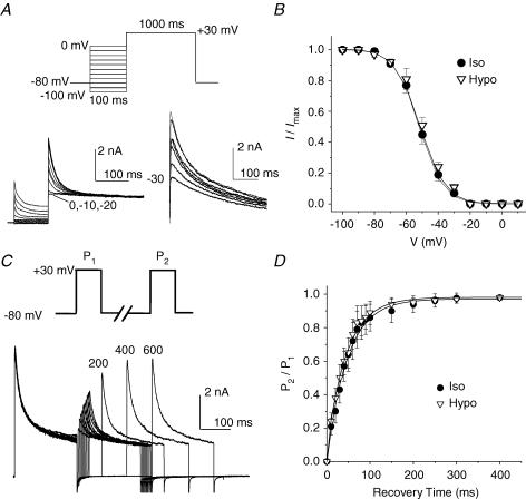
Figure 4. Effects of osmotic stress on voltage-dependent gating properties of Ito,fast in mouse ventricular apex myocytes.
A, the voltage dependence of steady-state inactivation of Ito,fast. Outward K+ currents were recorded during 1-s depolarization to +30 mV after 100-ms prepulses to potentials between −100 and 0 mV (protocol shown on the top) in the presence of 50 μm 4-AP. The representative current traces recorded under hyposmotic conditions are shown on the left. The test pulse currents obtained with the −20, −10 and 0 mV prepulses were superimposed on each other. The difference currents were obtained by subtraction of the test pulse currents recorded with the −20-mV prepulse from those recorded with prepulses between −100 and 0 mV. No inactivating component was observed with prepulses of −20, −10 and 0 mV. Numbers next to current traces indicate the corresponding potential of the inactivating prepulse. B, mean steady-state voltage-dependent inactivation curve of Ito,fast under isosmotic and hyposmotic conditions (n = 6). Peak Ito,fast recorded at +30 mV was normalized to the corresponding current amplitude measured at −100 mV. C and D, effects of cell swelling on the recovery from inactivation of Ito,fast; C, representative current traces recorded using a double-pulse voltage protocol (shown on the top); D, recovery of Ito,fast from inactivation under isosmotic and hyposmotic conditions (n = 7).

