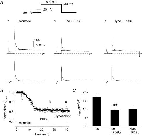
Figure 5. Effects of PKC activator (PDBu) on Ito,fast in mouse left ventricular apex myocytes.
A, representative current traces of Ito,fast (lower traces in each panel) obtained from the subtraction of currents recorded with the inactivating prepulse from those recorded without the inactivating prepulse (upper superimposed traces) in the presence of 50 μm 4-AP. Voltage protocols are shown on the top. The same cell was consecutively exposed to isosmotic solution (a), isosmotic + PDBu (100 nm, b), and hyposmotic + PDBu (100 nm, c). PDBu decreased Ito,fast under isosmotic conditions and subsequent hyposmotic perfusion failed to further activate Ito,fast. B, time course of changes in normalized Ito,fast at +30 mV when cells were exposed consecutively to isosmotic, isosmotic + PDBu, and hyposmotic + PDBu solutions. Peak Ito,fast was recorded every 1 min then normalized to the initial corresponding value at time 0 (n = 7). C, mean peak current densities of Ito,fast recorded when cells were exposed to isosmotic, isosmotic + PDBu, and hyposmotic + PDBu solutions. Currents were obtained using the protocols shown in panel A (n = 7). **P < 0.01 versus isosmotic (Iso) condition.

