
Figure 7.
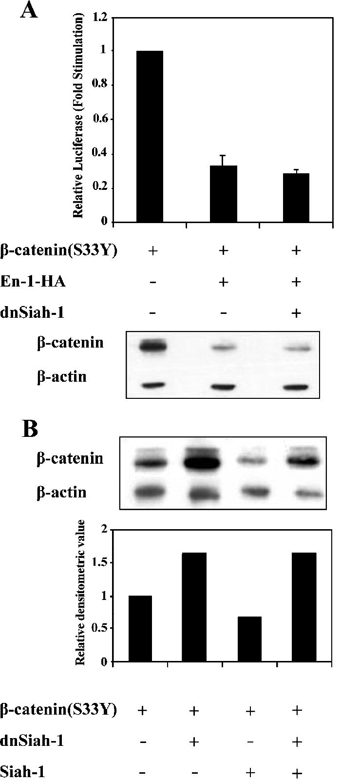
En-1–mediated β-catenin destabilization is Siah-1 independent. (A) The presence of dnSiah-1 does not affect En-1–repressive activity. 293T cells were transfected with β-catenin(S33Y) (0.1 μg), En-1-HA or empty vector (1 μg), dnSiah-1(1 μg) or empty vector, Cyclin D1/Luc (1 μg), and β-Gal (0.1 μg), and the luciferase and β-Gal levels were measured as described in the legend to Figure 1 (top). Cell lysates (30 μg) were subjected to Western analysis using anti-β-catenin or anti-β-actin antibody (bottom). (B) The presence of dnSiah-1 blocks Siah-1–mediated β-catenin degradation. 293T cells were transfected with β-catenin(S33Y) (0.1 μg), Siah-1 (0.1 μg) or empty vector, and dnSiah-1(1 μg) or empty vector. Cell lysates (30 μg) were subjected to Western analysis using anti-β-catenin or β-actin antibody (top). Densitometric analysis of the levels of β-catenin compared with β-actin levels is shown (bottom).