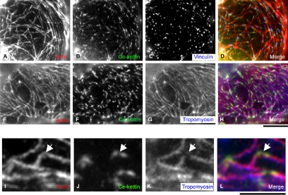
Figure 4.
Localization of Ce-kettin in ovarian nonstriated muscle. Myoepithelial sheath of the proximal ovary was stained for actin (A, E, and I), Ce-kettin (B, F, and J), and vinculin (C) or tropomyosin (G and K). Merged images of actin (red), Ce-kettin (green), and vinculin or tropomyosin (blue) are shown in D, H, and L. High-magnification images in I–L show a representative area where differential localization of Ce-kettin and tropomyosin is clearly observed (arrows). Bars, 10 μm (A–H) and 5 μm (I–L).

