Figure 5.
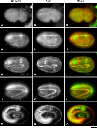
Localization of Ce-kettin in embryos. Embryos at the comma (A–C), twofold (D–I), and threefold (J–O) stages were stained for Ce-kettin (A, D, G, J, and M) and actin (B, E, H, K, and N). Merged images of Ce-kettin (green) and actin (red) are shown in C, F, I, L, and O. Focus was adjusted for the body wall muscle in D–F and J–L or the pharynx in G–I and M–O. Actin-rich structure in H (asterisk) is the nerve ring. Bar, 10 μm.

