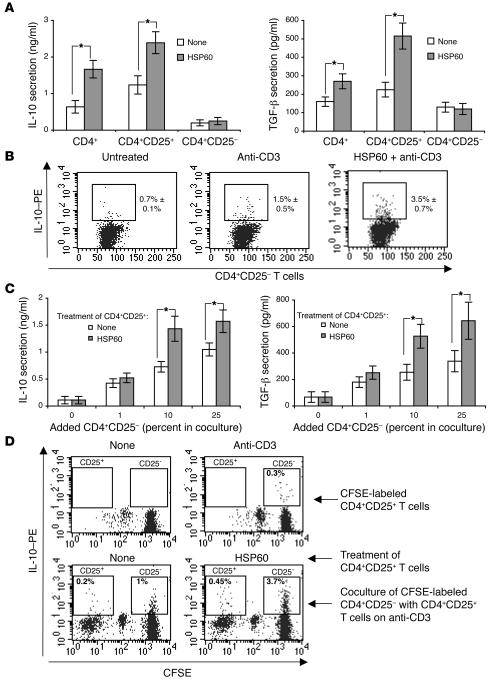
Figure 7. HSP60 induces IL-10 and TGF-β secretion in CD4+ CD25+ T cells.
Unseparated CD4+ (A), CD4+CD25+ (A–D), or CD4+CD25– T cells (A) were incubated with HSP60 (1 ng/ml) for 2 hours, washed, and transferred to 24-well plates coated with anti-CD3 mAb (OKT; 0.5 μg/ml) separately (A and B), or cocultured in the indicated proportions (C and D) in serum-free medium. IL-10 and TGF-β secretion was analyzed after 24 hours by ELISA (A and C), or by FACS (B and D). (D) CD4+CD25– T cells were labeled with CFSE and washed before coculture with CD4+CD25+ T cells. (A and C) The means ± SD of 5 different donors are shown. *P < 0.05. (B and D) FACS histograms are representative of 3 different donors. Numbers indicate average percentage ± SD of 3 different donors.

