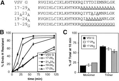
Figure 6.
Secondary structure of the tail may substitute for specific amino acid sequence. (A) Amino acid sequence (single-letter code) of wild-type and mutant VSV G proteins. Substituted residues are underlined. (B) Kinetics of acquisition of endo H resistance of VSV G, 17–29A, 21A23A, 19–24A, and 17–29G. BHK-21 cells expressing these proteins were pulse labeled for 5 min and chased for the times indicated. Immunoprecipitated proteins were subjected to endo H treatment as described in MATERIALS AND METHODS and separated by SDS-PAGE. The amount of endo H-resistant protein was quantitated by phosphorimaging. Each time point represents the mean of at least three experiments ± SD. (C) Oligomerization of VSV G, 17–29A, and 17–29G. BHK-21 cells expressing VSV G, 17–29A, or 17–29G were pulse labeled for 5 min and solubilized after a 10-min chase. Lysates were loaded onto 5–20% linear sucrose gradients and centrifuged as described in MATERIALS AND METHODS. Fractions were collected, immunoprecipitated with anti-VSV antibody, and analyzed by SDS-PAGE. The percentage of protein found as monomer (4S) or trimer (8S) relative to the total pool of VSV G or mutant proteins was quantitated using phosphorimager analysis. Each time point represents the mean of two experiments ± SEM.

