Figure 2.
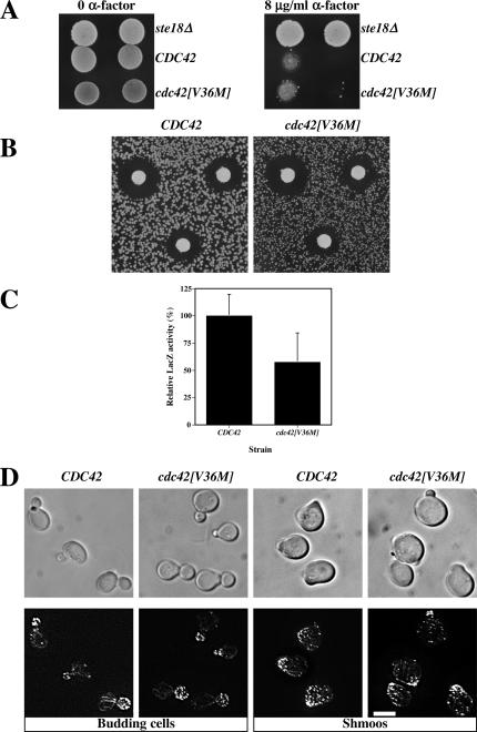
cdc42[V36M] cells respond to mating pheromone. (A) cdc42[V36M] cells are not resistant to mating pheromone. CDC42 (RAY1772), cdc42[V36M] (RAY1926), and ste18Δ strains were spotted on YEPD plates containing or lacking 8 μg/ml α-factor and incubated for 5 d. (B) cdc42[V36M] cells arrest growth in the presence of mating pheromone similar to wild-type cells. α-factor (1, 0.5, and 0.2 μg) was spotted on filters placed on a lawn of the indicated strain. Plates were incubated for 3 d. Measurements of the halo diameter indicated ≤5% difference between CDC42 and cdc42[V36M] halos. (C) cdc42[V36M] cells induce the mating-specific FUS1 gene in a pheromone-dependent manner. Cells containing the FUS1-lacZ plasmid pSG231 were incubated with 10 μM α-factor for 1 h, and LacZ activity was determined. The means of four determinations from two independent experiments are shown. Error bars, SD. LacZ activity for CDC42 cells (21.0 Miller units) was set at 100%. (D) cdc42[V36M] cells polarize their actin cytoskeleton. DIC and fluorescence images of budding cells and cells treated with 12 μM α-factor for 2 h are shown. Actin cytoskeleton is visualized with Alexa-488 phalloidin. Fluorescence images are maximum intensity projections of Z-sections (8–12 × 0.1 μm).

