Figure 4.
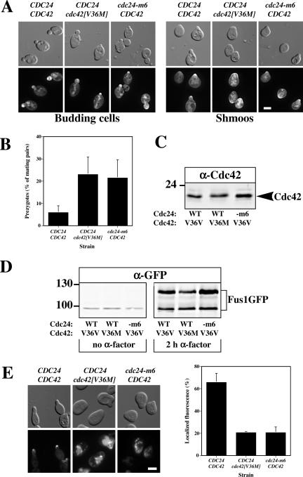
Cdc42p and Cdc24p are required for correct localization of the cell fusion protein Fus1. (A) GFP-Cdc42[V36M] localizes to the bud and shmoo tips. Indicated strains (RAY1830, RAY1951, and RAY1833), which are MATa cdc24Δ cdc42Δ cells carrying a plasmid copy of CDC24 and GFP-Cdc42 or CDC24 and GFP-Cdc42[V36M] or cdc24-m6 and GFP-Cdc42, each behind their respective endogenous promoter on a CEN plasmid, were either grown in the absence (left panels) or presence (right panels) of 12 μM α-factor for 2 h, and fluorescence and DIC images were taken. (B) cdc24-m6 and cdc42[V36M] mating mixtures accumulate similar amounts of prezygotes. Indicated strains (RAY1793, RAY1801, and RAY1952) were mated with RAY1487 (GFP-Bud1) and analyzed as in 3D. Values are the means of four independent matings (n = 200 mating pairs). Error bars, SD. (C) Expression levels of Cdc42p in CDC42/CDC24, cdc42[V36M]/CDC24, and CDC42/cdc24-m6 cells. Extracts of logarithmically growing cells (RAY1793, RAY1801, and RAY1952) were analyzed by immunoblot and probed with anti-Cdc42p polyclonal sera. Similar results were observed in three independent experiments. (D) cdc42[V36M] cells induce Fus1-GFP in the presence of mating pheromone. Extracts of cells from the same experiment as above were analyzed by immunoblot either in the absence or presence of α-factor. The lower band is an unglycosylated form of Fus1-GFP. Similar results were observed in three independent experiments. (E) Fus1-GFP is not correctly localized in cdc24-m6 and cdc42[V36M] shmoos. Indicated strains (RAY1793, RAY1801, and RAY1952) carrying a Fus1-GFP plasmid were incubated with 12 μM α-factor for 2 h, and fluorescence images were taken. The mean percentage of shmoos with an observable fluorescence signal at the tip of the mating projection was determined from two independent experiments (n = 200). Bars indicate values.

