Abstract
The identification and characterization of microparticles has become an important field of study in recent years due to their presence in the environment and association with pathogenesis. Asbestos fibers have been intensively studied for these reasons. Since conventional microscopy has not provided unique identification of these materials, electron probe microanalysis, which yields chemical data, has been utilized in conjunction with other techniques to provide the necessary answers.
The options now available to undertake electron probe analysis are discussed with relation to their utilization for microparticle analyses. Two types of electron sources are available, thermionic and field emission. The x-ray spectroscopy requires the use of either wavelength-dispersive focussing crystal spectrometers or an energy-dispersive Si(Li) x-ray detector.
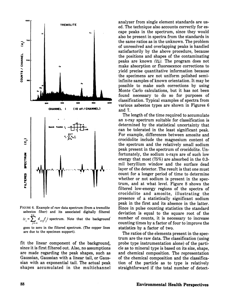
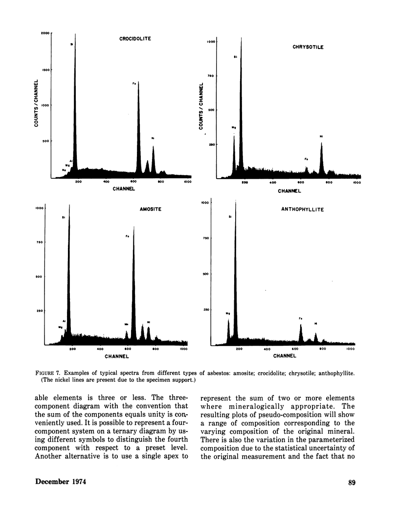
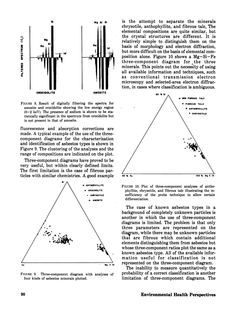
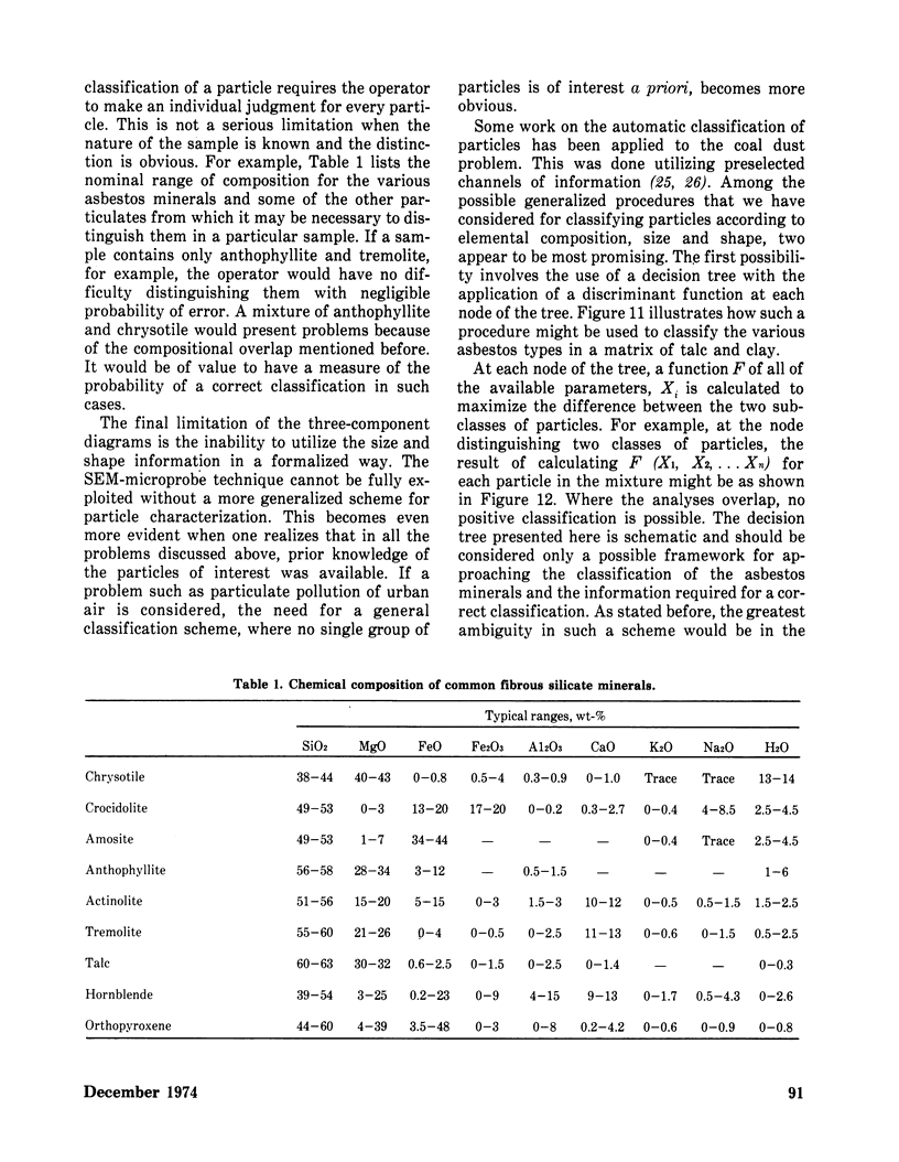
Data are presented to demonstrate the feasibility of asbestos identification by using modified raw data obtained with a scanning electron microscope and energy-dispersive x-ray spectrometer. Further, the extension of the technique to other microparticle identification problems is discussed.
Full text
PDF
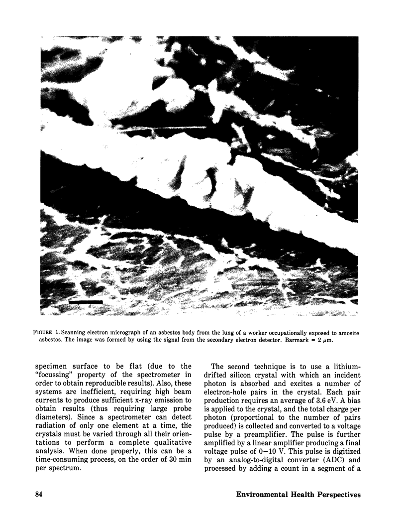
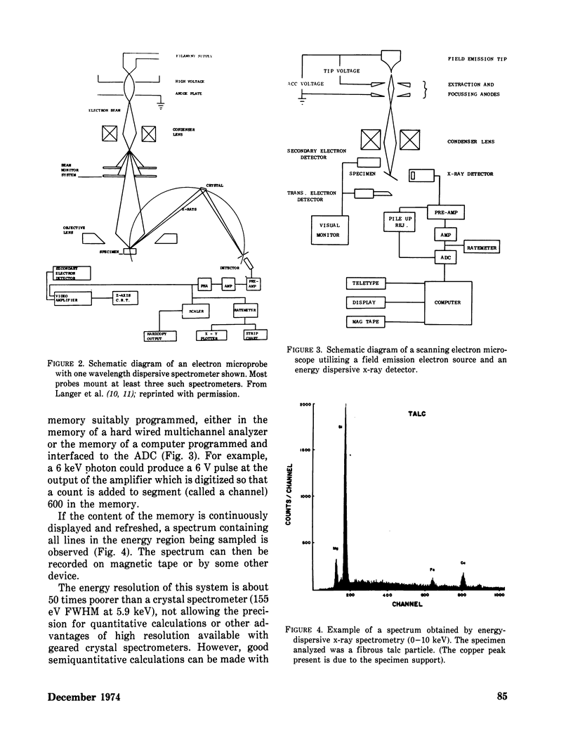
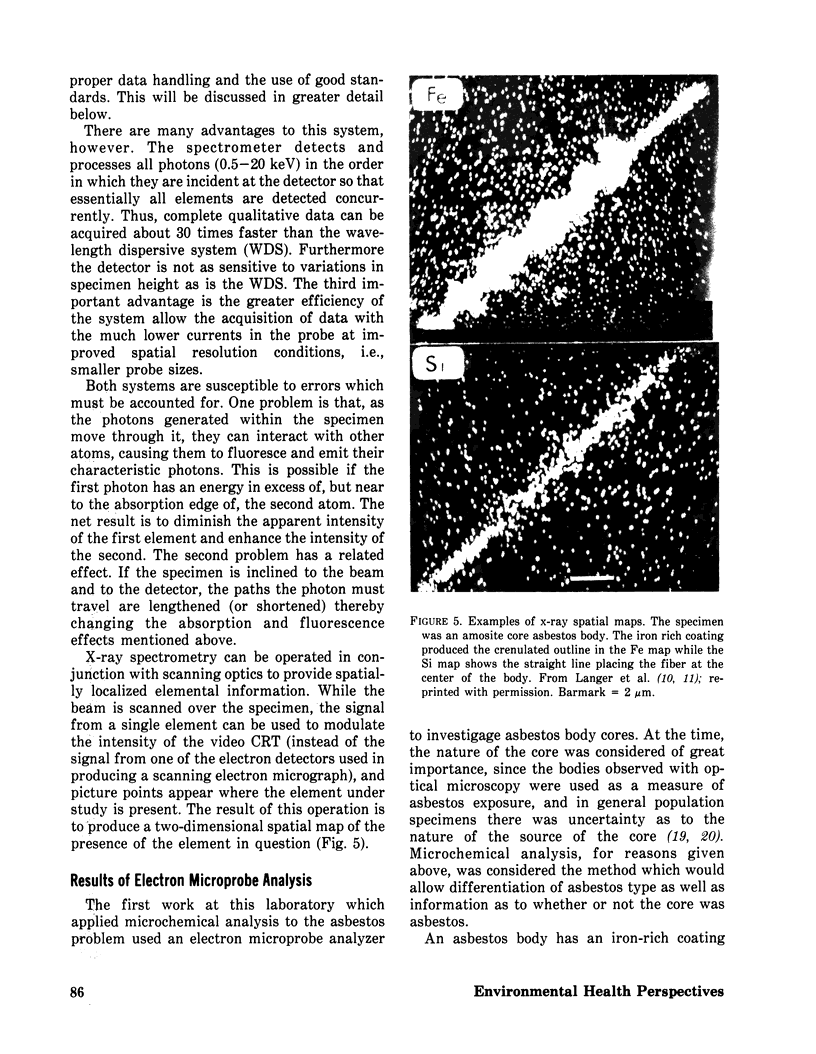
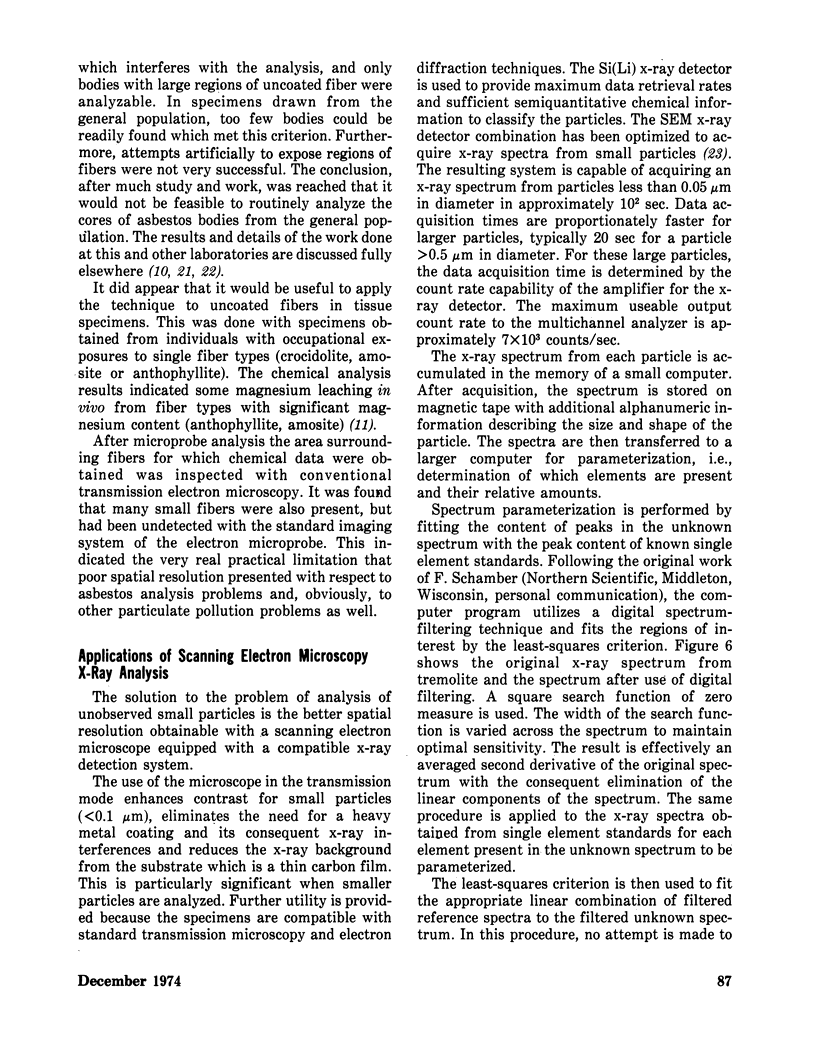
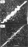
Images in this article
Selected References
These references are in PubMed. This may not be the complete list of references from this article.
- Andersen C. A. An introduction to the electron probe microanalyzer and its application to biochemistry. Methods Biochem Anal. 1967;15:147–270. doi: 10.1002/9780470110331.ch4. [DOI] [PubMed] [Google Scholar]
- Langer A. M., Mackler A. D., Pooley F. D. Electron microscopical investigation of asbestos fibers. Environ Health Perspect. 1974 Dec;9:63–80. doi: 10.1289/ehp.74963. [DOI] [PMC free article] [PubMed] [Google Scholar]
- Langer A. M., Rubin I. B., Selikoff I. J. Chemical characterization of asbestos body cores by electron microprobe analysis. J Histochem Cytochem. 1972 Sep;20(9):723–734. doi: 10.1177/20.9.723. [DOI] [PubMed] [Google Scholar]
- Langer A. M., Rubin I. B., Selikoff I. J., Pooley F. D. Chemical characterization of uncoated asbestos fibers from the lungs of asbestos workers by electron microprobe analysis. J Histochem Cytochem. 1972 Sep;20(9):735–740. doi: 10.1177/20.9.735. [DOI] [PubMed] [Google Scholar]
- SELIKOFF I. J., CHURG J., HAMMOND E. C. ASBESTOS EXPOSURE AND NEOPLASIA. JAMA. 1964 Apr 6;188:22–26. doi: 10.1001/jama.1964.03060270028006. [DOI] [PubMed] [Google Scholar]
- Selikoff I. J., Hammond E. C., Churg J. Carcinogenicity of amosite asbestos. Arch Environ Health. 1972 Sep;25(3):183–186. doi: 10.1080/00039896.1972.10666158. [DOI] [PubMed] [Google Scholar]
- Skikne M. I., Talbot J. H., Rendall R. E. Electron diffraction patterns of U.I.C.C. asbestos samples. Environ Res. 1971 Apr;4(2):141–145. doi: 10.1016/0013-9351(71)90042-9. [DOI] [PubMed] [Google Scholar]
- Speil S., Leineweber J. P. Asbestos minerals in modern technology. Environ Res. 1969 Apr;2(3):166–208. doi: 10.1016/0013-9351(69)90036-x. [DOI] [PubMed] [Google Scholar]
- WAGNER J. C., SLEGGS C. A., MARCHAND P. Diffuse pleural mesothelioma and asbestos exposure in the North Western Cape Province. Br J Ind Med. 1960 Oct;17:260–271. doi: 10.1136/oem.17.4.260. [DOI] [PMC free article] [PubMed] [Google Scholar]


