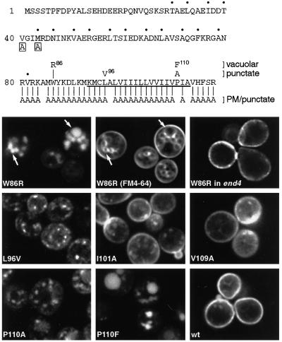
Figure 4.
GFP-Snc1p point mutants. At the top the sequence of Snc1p is shown. Dots indicate the heptad repeat involved in SNARE complex formation, and the transmembrane domain is underlined. Boxed letters show the point mutations that affect endocytosis. For residues 80–117, the mutations and the distributions of the mutant proteins are summarized. All but one of the Ala substitutions gave plasma membrane staining, with variable amounts of punctate staining in addition. The lower panels show vacuolar staining of W96R in wild type (with FM4-64 double label) but not in end4 cells, and the patterns in wild-type cells of L96V, P110A, P110F, and two examples of the more variable Ala substitutions (I101A and V109A). Unmutated GFP-Snc1p (wt) was analyzed in parallel for comparison. The FM4-64-labeled cells were photographed in the presence of the dye and thus show plasma membrane as well as vacuolar staining.

