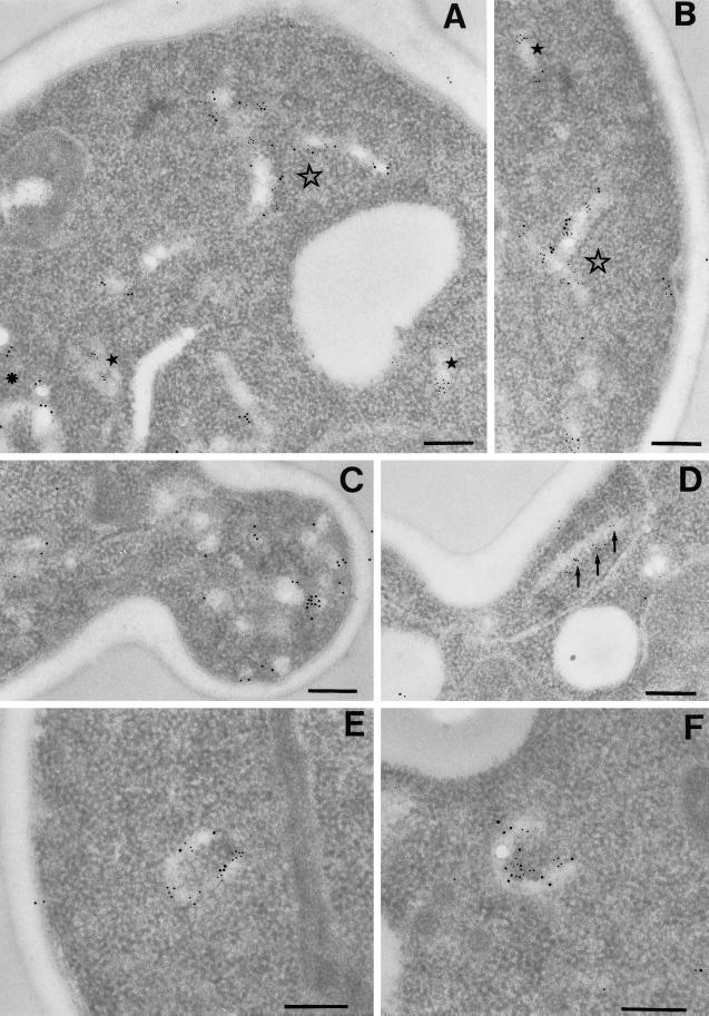
Figure 9.
Immuno-EM of SNAREs. Immunolocalization of SNAREs on thin sections was performed as described in MATERIALS AND METHODS. Pep12p and Sed5p were detected using 5 nm IgG-colloidal gold, and Tlg1p was detected using 10 nm IgG-colloidal gold. The contrast of the gold particles has been digitally enhanced for clarity. (A and B) Localization of Pep12p and Tlg1p. Filled stars denote structures with only Pep12p labeling; the asterisk marks structures with only Tlg1p labeling; and the unfilled star marks structures showing colocalization of Pep12p and Tlg1p. (C–F) Localization of Sed5p and Tlg1p. The arrows mark a structure labeled only with Sed5p. Note the 100-nm vesicles with only Tlg1p labeling in C and structures showing colocalization of Sed5p and Tlg1p in E and F. Bars, 200 nm.

