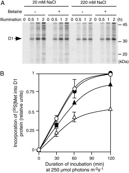
Figure 5.
Betaine accelerated de novo synthesis of D1 protein during photoinhibition under salt stress. PAMCOD cells were labeled with [35S]Met during incubation in light at 250 μmol photons m−2 s−1 as described in “Materials and Methods.” Thylakoid membranes were isolated from cells that had been harvested at designated times and proteins in thylakoid membranes were solubilized and separated by SDS-PAGE as described in “Materials and Methods.” A, Autoradiogram. B, Time course of changes in levels of D1 protein that had been labeled with [35S]Met during incubation in light at 250 μmol photons m−2 s−1. ○, Control cells (20 mm NaCl); •, choline-supplemented cells (20 mm NaCl); ▵, control cells (220 mm NaCl); ▴, choline-supplemented cells (220 mm NaCl). Each point and error bar represents the average and sd, respectively, of results from three independent experiments.

