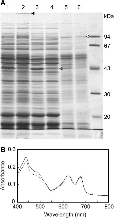
Figure 6.
Comparison of the polypeptide composition and the absorption spectrum between wild-type and Δ1848 Δ2060 cells. A, SDS-PAGE of cell-free lysate fractions (lane 1, wild type; lane 2, Δ1848 Δ2060), soluble fractions (lane 3, wild type; lane 4, Δ1848 Δ2060), and membrane fractions (lane 5, wild type; lane 6, Δ1848 Δ2060) on a 10% polyacrylamide gel. Cell-free lysates and membrane fractions equivalent to 2 μg Chl and the corresponding amounts of soluble fractions were applied. The gel was stained with Coomassie Brilliant Blue R-250. B, Absorption spectra for wild-type (line) and Δ1848 Δ2060 (broken line) cells. Wild-type and Δ1848 Δ2060 cells were harvested after 96 h at 30°C at OD730 values of 2.15 and 1.94, respectively. Cells were diluted to an OD730 value of 0.05 before measuring the spectra.

