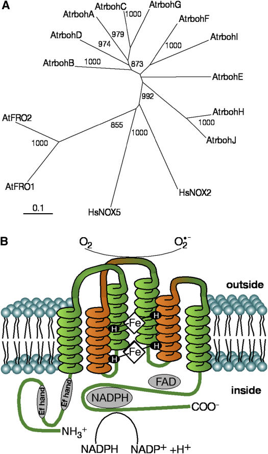
Figure 1.
Structure of Rboh and their phylogenetic distribution. A, Phylogenetic tree comparing Arabidopsis Atrboh with AtFRO and mammalian HsNOX2 and HsNOX5 proteins. Atrboh members are listed in Table I. Comparisons included HsNOX2 (P04839), HsNOX5 (AF317889), AtFRO1 (At1g01590), and AtFRO2 (At1g01580). Only the carboxy terminus with homology to gp91phox/NOX2 (excluding the EF hands) was used in the alignment. The phylogenetic analyses were made by neighbor-joining tree with ClustalX. The length of the horizontal lines connecting the sequences is proportional to the estimated amino acid substitutions/site between these sequences. Bootstrap values from 1,000 iterations are shown. B, Schematic diagram of Rboh structure as predicted to be located in the membrane, showing the irreversible transfer of charge from cellular NADPH to EC oxygen. Shown are the N-terminal region EF hands juxtaposed to the C-terminal end to indicate an interaction by which calcium-dependent activity is regulated.

