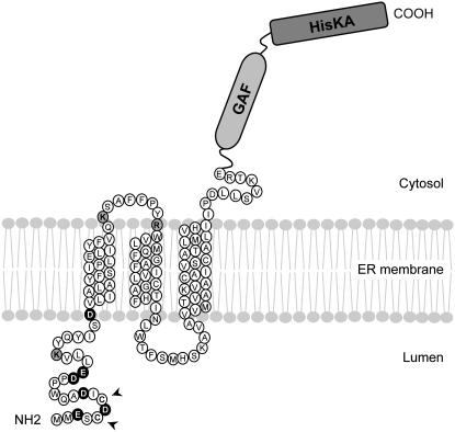
Figure 7.
A proposed model for CmERS1 topology on the ER membrane. Based on membrane fractionation, GFP imaging, protease protection assay, and glycosylation analysis, we propose that CmERS1 is predominantly localized to the ER and spans the membrane three times with its N terminus facing the luminal space and its C terminus lying on the cytosolic side. The positively and negatively charged residues in TM1-flanking regions are shaded gray and black, respectively. Arrows indicate the residues that are possibly involved in disulfide-linked dimerization of the receptor. The TM helices were identified by PHD 2.1 (Rost et al., 1996).

