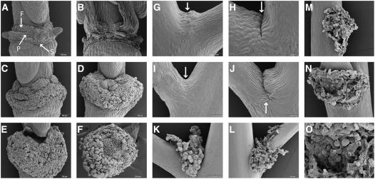
Figure 2.
Scanning Electron Micrographs of Floral AZs and the Base of the Pedicel.
(A) Wild-type AZ of sepals (S), petals (P), and filaments (F) after abscission, as indicated.
(B) Mutant ida AZ with broken cells after forcible removal of floral organs.
(C) Enlarged 35S:IDA AZs.
(D) 35S:IDA AZ with a dramatically greater number of rounded cells.
(E) Gynophore detached from the 35S:IDA AZ.
(F) 35S:IDA AZ showing broken cells where the gynophore was attached.
(G) Wild-type pedicel vestigial AZ with a band of small cells (arrow).
(H) Wild-type pedicel vestigial AZ. A small cleft (arrow) developed in older pedicels.
(I) and (J) Pedicel AZs in 35S:IDA plants at stages comparable to the wild type shown in (G) and (H).
(K) and (L) Pedicel AZs in 35S:IDA plants, later stages.
(M) Base of the pedicel in a 35S:IDA plant showing rounded cells after abscission of the pedicel.
(N) AZ showing rounded cells after shedding of the cauline leaf in a 35S:IDA plant.
(O) Close-up of the cells in the cauline leaf AZ.

