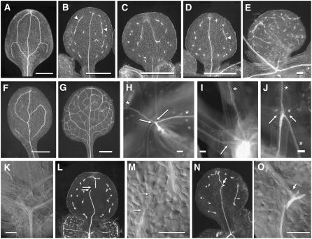
Figure 4.
Loss of AGD1, AGD2, and AGD4 Enhances sfc Vein Pattern Defects.
(A) Wild-type cotyledon vein pattern.
(B) sfc-9 cotyledon vein pattern. Arrowheads point to elongated VIs.
(C) agd1 agd2 agd4 sfc-9 quadruple mutant cotyledon. Note the loss of primary vein and small sized VI.
(D) agd1 agd2 agd4 sfc-9 quadruple mutant cotyledon; approximately half retain an intact primary vein. Arrowhead points to uncommon elongated VI.
(E) agd1 agd2 agd4 sfc-9 quadruple mutant leaf; compare with sfc leaf vein patterns in Figure 1. This leaf shows greater interruption of marginal vein network and smaller VIs than in sfc single mutants.
(F) agd1 agd2 adg4 triple mutant cotyledons occasionally had incomplete proximal and distal areoles.
(G) agd1 agd2 agd4 triple mutant leaf vein pattern is similar to the wild type.
(H) to (J) Connection of cotyledon and leaf primary veins with the stem/hypocotyl vasculature. Asterisks indicate cotyledon veins, and arrows point to the junctions between leaf primary veins and the stem.
(H) sfc-9 leaf veins always connect with the stem's vascular system.
(I) agd1 agd2 agd4 triple mutant leaf veins always connect to the stem's vascular system.
(J) agd1 agd2 agd4 sfc-9 quadruple mutant leaf veins failed to connect to the stem's vascular system.
(K) DIC image of agd1 ad2 agd4 sfc-9 quadruple mutant leaf vein base shows neither vascular nor procambial connections between the leaf primary vein and the hypocotyl.
(L) and (M) agd1 agd2 agd4 sfc-9 quadruple mutant with interrupted primary vein shown in dark field (L) and the region of primary vein disruption (M). Arrows indicate the extent of procambium that extended above the lower segment of the primary vein.
(N) and (O) Another agd1 agd2 agd4 sfc-9 quadruple mutant showing a zigzag primary vein (arrows).
Bars = 1 mm ([A] to [D], [F], and [G]) and 100 μm ([E], [H] to [K], [M], and [O]).

