Figure 6.
DR5:GUS Expression and Root Development in sfc Mutants.
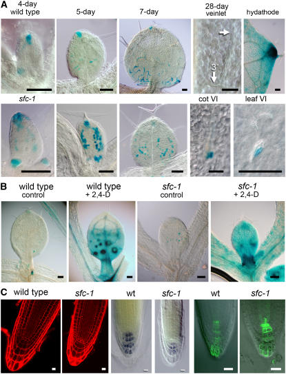
(A) DR5:GUS expression in developing leaves of the wild type (top row) and sfc-1 mutant (bottom row). From the left are representative day 4, day 5, and day 7 leaves. The right two images of both rows show tissue from a 28-d-old plant. In the wild type (top), a leaf from a 28-d-old plant no longer has GUS staining associated with differentiated veins (arrow shows veinlet; 3° refers to a tertiary vein), but the same leaf shows strong GUS staining associated with hydathodes. In 28-d-old sfc-1 mutants, DR5:GUS staining is maintained in cells associated with VIs of both cotyledons and leaves. Bars = 0.1 mm.
(B) A 5-h treatment with 5 μm 2,4-D induces DR5:GUS staining in both the wild type and sfc mutants. Large spots in the wild type correspond to differentiating trichomes. Bars = 0.1 mm.
(C) Roots of the wild type and sfc mutants have similar cellular organization (propidium iodide–stained 6-d-old tissue to the far left), although the sfc cells are smaller and the root has a more blunt shape. Starch staining shows similar patterns of cell types (center, 6-d-old seedling), and DR5(rev):GFP (right, 6-d-old seedling) shows similar expression patterns. Bars = 10 μm.

