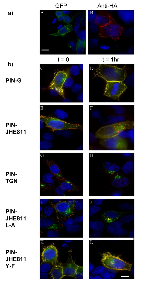
Figure 6.

a) Total versus surface expression of pIN-JHE811 – a fusion construct corresponding to pIN-G bearing the cytoplasmic carboxy terminus of Kv1.4. HEK293 cells transiently transfected with pIN-JHE811 were fixed, stained and imaged on a Delta-Vision workstation. Panel A demonstrates total cellular pIN-JHE811 fluorescence (green) whilst panel B depicts surface staining of pIN-JHE811 with anti-HA antibodies (red: as described in Methods). Blue indicates DAPI-stained nuclei. b) Site-directed mutagenesis of pIN-JHE811 reveals internalization signals in the Kv1.4 carboxy terminus. HEK293 cells transiently transfected with pIN-G (panels C and D), pIN-JHE811 (panels E and F), pIN-TGN (panels G and H), pIN-JHE811 L-A (panels I and J) and pIN-JHE811 Y-F (panels K and L) and were labeled with anti-HA antibodies and either fixed directly (panels C, E, G, I and K) or incubated for 1 hour at 37°C to allow antibody internalization (panels D, F, H, J and L). Internalized antibody was then detected using Cy3-conjugated 2° goat anti-mouse antibody. Green denotes PIN-protein fluorescence while red corresponds to the anti-HA antibody. Areas of red/green overlap are shown in yellow. Blue indicates DAPI-stained nuclei. Scale Bar: 15 μM.
