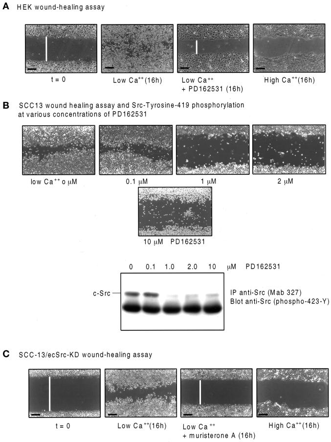
Figure 6.
The Src-inhibitory drug PD162531 and overexpression of the dominant-negative c-Src protein suppress keratinocyte wound healing in vitro. Phase-contrast photomicrographs show four individual HEK monolayers (A) or SCC-13/ecSrc-KD cells (B and C) grown to confluence in low Ca2+ and then wounded (example shown at t = 0) and either treated for 16 h (16h) with 0.02% DMSO (low Ca2+, 0 μM), 0.1, 1, 2, or 10 μM PD162531 as indicated (low Ca2+ + PD162531), or 10 μM muristerone A [low Ca2+ muristerone A (16h)] or transferred to high Ca2+ for 16 h [high Ca2+ (16h)]. Bars, 200 μm. White bars represent relative distances between the two edges of apposing epithelial sheets. Presumed c-Src autophosphorylation at the indicated drug concentrations was monitored by immunoprecipitating cell lysates with anti-Src (mAb 327) and immunoblotting using anti-Src (phospho-423-Y) as probe (B).

