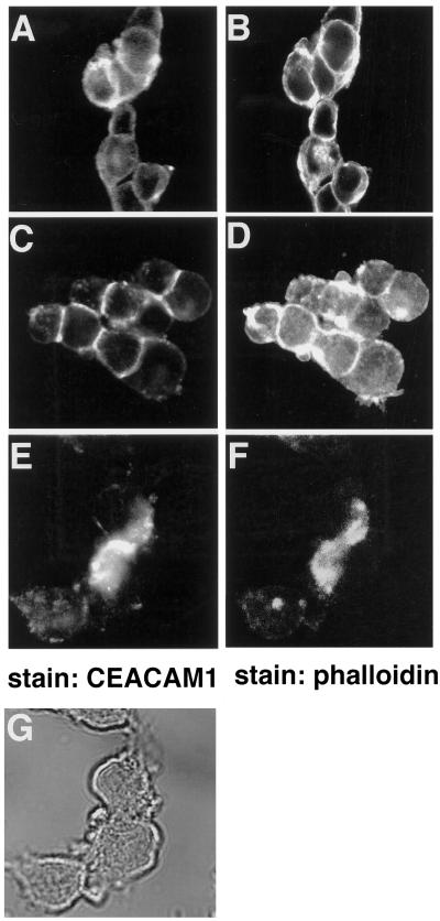
Figure 4.
Localization of the CEACAM1 isoforms in CT51 intestinal cells. CT51 cells stably expressing either the CEACAM1-S (A and B) or CEACAM1-L (C–G) were either untreated (A–D) or treated (E–G) with 1 μM of the cytochalasin D for 1 h and processed for indirect immunofluorescence. CEACAM1 was detected with the CC1 monoclonal antibody and FITC-labeled rabbit anti-mouse secondary antibodies (A, C, and E). Actin in the same cells was revealed using TRITC-conjugated phalloidin (B, D, and F). (G) Phase contrast of cells in E and F.

