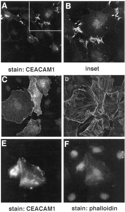
Figure 8.
Localization of CEACAM1-L at cell–cell boundaries is inhibited by treatment with anti-CEACAM1 antibodies or cytochalasin D treatment. Quiescent serum-starved Swiss 3T3 fibroblasts were injected with a pXM139 vector expressing CEACAM1-L and a pRK5 vector encoding the myc-tagged constitutively activated L61Cdc42 protein. The cells were treated for 6 h with 250 ng/ml Fab fragments of the anti-CEACAM1 231 polyclonal antibody (C and D) or with Fab fragments of the preimmune serum (A and B) added 2 h after the injections. After 10 h of expression, permeabilized cells were stained for CEACAM1-L expression using the 231 anti-CEACAM1 polyclonal antibody and an FITC-conjugated anti-rabbit antibody (A, C, and E), and actin was detected using TRITC-conjugated phalloidin (B, D, and F). In E and F, 2 h after microinjections, the microinjected cells were treated with cytochalasin D (50 ng/ml) for 6 h and were processed for immunofluorescence at the end of this treatment, as described above. Expression of CEACAM1-L at cell–cell boundaries was not detected, indicating that association with the actin cytoskeleton is necessary for CEACAM1-L localization at sites of cell–cell contact.

