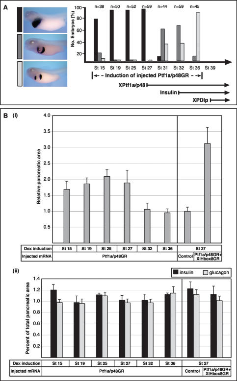
Figure 3.
The Ptf1a/p48-mediated increase of ectopic exocrine and late endocrine cells requires uncommitted endoderm. Dexamethasone induction of injected embryos was performed at different time points in between stage 15 and stage 36, as indicated. The effects on pancreas development were evaluated by whole-mount in situ hybridization analysis of XPDIp expression at stage 41 for the exocrine pancreas, and double-staining immunohistochemical analysis of insulin and glucagon expression at stage 48 for the endocrine pancreas. For morphometric quantification, the pixel number of insulin-positive and glucagon-positive cells was measured separately in serial sections using Adobe. (A) The extent of ectopic XPDIp expression at stage 41 is dependent on the stage of induction of injected Ptf1a/p48GR. The bottom right part illustrates the temporal expression profile of endogenous XPtf1a/p48, insulin, and XPDIp. (B) Despite an increase in total pancreatic area, there is no significant difference in the ratio of endocrine to exocrine pancreatic cells in embryos overexpressing Ptf1a/p48 alone or in combination with XlHbox8. (Panel i) Ectopic expression of Ptf1a/p48GR alone or in combination with XlHbox8GR results in a roughly two- and threefold increase in total pancreatic area relative to uninjected control embryos, respectively. (Panel ii) No significant difference is observed for the ratio of endocrine to total pancreatic area in a comparison of different time points for dexamethasone treatment. Sectioned pancreatic tissue was immunostained for insulin and glucagon, nuclei were counterstained with DAPI. Boundaries of the pancreatic area were delineated on the basis of morphology and pixel quantification was performed using Adobe Photoshop. An entire series of sections was analyzed for eight different embryos in each experiment. The average total pixel number of a series of pancreatic sections from control embryos is referred to as 1.

