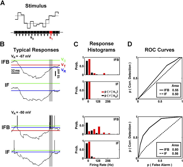
Figure 4. Detection of the Offset of Inhibitory Luminance Sequences.
(A) LGN responses to a noisy stimulus in which an inhibitory sequence randomly appeared were simulated. The stimulus was classified as S 0 (black) or S 1 (red) depending on whether or not each interval contained the excitatory transient of the sequence. A typical realization of the stimulus with SNR = 1/2 and sequence duration = 128 ms is shown (intensity averaged over all pixels in RF center). The black line indicates the actual stimulus and the gray line indicates the underlying sequence.
(B) Voltage traces of the IFB and IF responses to the stimulus shown in (A) at two different resting potentials, V R = −67 mV (top) and V R = −50 mV (bottom), with V T = −60 mV. The interval in the response that corresponds to condition S 1 is shaded (response was shifted for presentation to remove latency between stimulus and response). The spike threshold ( V Θ, green), burst de-inactivation potential and threshold ( V T, red), and resting potential ( V R, blue) are shown.
(C) The probability distributions of the firing rate of the IFB and IF models during the S 0 (black) and S 1 (red) stimulus conditions at V R = −67 mV (top) and V R = −50 mV (bottom).
(D) ROC curves for the IFB and IF models at V R = −67 mV (top) and V R = −50 mV (bottom). The area under the ROC curve is indicated.

