
Table 1.
Details of medial thalamus lesions in all rats, expressed as percentages of bilateral volumes
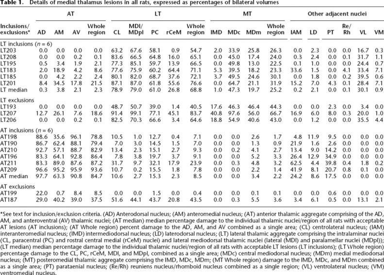
*See text for inclusion/exclusion criteria. (AD) Anterodorsal nucleus; (AM) anteromedial nucleus; (AT) anterior thalamic aggregate comprising of the AD, AM, and anteroventral (AV) thalamic nuclei; (AT median) median percentage damage to the individual thalamic nuclei/region of all rats with acceptable AT lesions (AT inclusions); (AT Whole region) percent damage to the AD, AM, and AV combined as a single area; (CL) centrolateral nucleus; (IAM) interanteromedial nucleus; (IMD) intermediodorsal nucleus; (LD) laterodorsal nucleus; (LT) lateral thalamic aggregate comprising the intralaminar nuclei (CL, paracentral (PC) and rostral central medial (rCeM) nuclei) and lateral mediodorsal thalamic nuclei (lateral (MDl) and paralamellar nuclei (MDpl)); (LT median) median percentage damage to the individual thalamic nuclei/region of all rats with acceptable LT lesions (LT inclusions); (LT Whole region) percentage damage to the CL, PC, rCeM, MDl, and MDpl, combined as a single area; (MDc) central mediodorsal nucleus; (MDm) medial mediodorsal nucleus; (MT) posteromedial thalamic aggregate comprising the IMD, MDc, MDm; (MT Whole region) damage to the IMD, MDc, and MDm combined as a single area; (PT) parataenial nucleus; (Re/Rh) reuniens nucleus/rhomboid nucleus combined as a single region; (VL) ventrolateral nucleus; (VM) ventromedial nucleus.