
Figure 2.
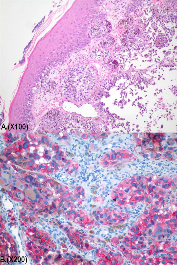
Tissue sections revealed extensive infiltration of the mucosa by neoplastic predominantly epithelioid cells, in a solid, nested, trabecular or alveolar pattern. Continuity of the tumor with the surface epithelium was identified (Haematoxylin and Eosin stain, magnification ×100) (A). The neoplastic cells demonstrate strong immunoreactivity for Melan A monoclonal antibody (magnification ×200) (B).